

What the study found
Craniofacial osteomas were most commonly located in the frontal bone, and females made up most cases. The study also found no pathogenic variants related to Gardner syndrome in the patients who underwent genetic testing, and no recurrences were seen during 6 months of follow-up.
Why the authors say this matters
The authors conclude that combining imaging-based diagnosis, tailored surgery, and selective genetic testing allows for accurate evaluation, effective treatment, and favorable postoperative outcomes. They also state that the study helps establish a systematic approach for diagnosing, evaluating, and managing craniofacial osteomas.
What the researchers tested
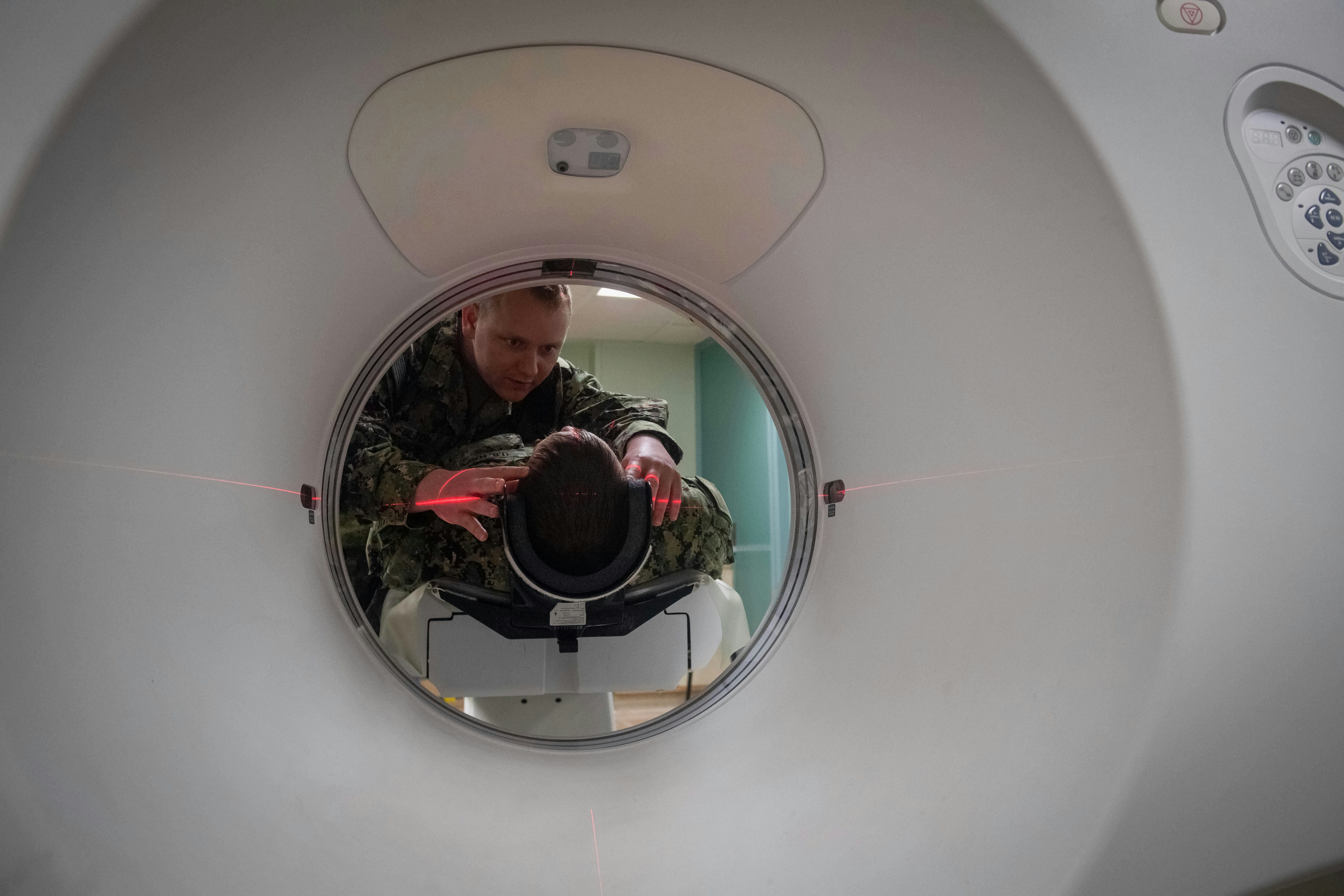
The researchers retrospectively reviewed 141 patients with craniofacial osteomas treated at one hospital between October 2011 and September 2025. All patients had clinical examinations and 3-dimensional computed tomography, and surgery was done with direct, endoscopic, or bicoronal approaches depending on lesion characteristics. Whole exome sequencing, a broad genetic test that examines many genes at once, was used in patients with multiple large osteomas to look for changes in EXT1, EXT2, APC, MSH2, and MLH1.
What worked and what didn't
A total of 148 osteomas were identified. The frontal bone accounted for 60.1% of lesions, followed by the parietal, mandibular, and occipital bones, and females represented 79.1% of cases. Genetic testing did not identify pathogenic variants related to Gardner syndrome, and no recurrences were observed during the 6-month follow-up period.
What to keep in mind
The abstract does not describe detailed limitations beyond the retrospective, single-hospital design and the 6-month follow-up period. Genetic testing was selective rather than performed in all patients, so the genetic findings apply only to the tested subgroup.
Key points
- Craniofacial osteomas were most often found in the frontal bone.
- Females accounted for 79.1% of the cases in this series.
- Selective whole exome sequencing found no pathogenic variants related to Gardner syndrome.
- No recurrences were observed during 6 months of follow-up.
- The study used clinical examination, 3-dimensional CT, and tailored surgical approaches.
Disclosure
- Research title:
- Craniofacial osteomas were most often found in the frontal bone
- Authors:
- Jung-Eun Moon, Hyun Su Kang, Yong June Chang, Ki-Su Park, Mansoo Suh, Jeong Yeop Ryu, Kang Young Cho, Jung Dug Yang, Ho Yun Chung, Joon Seok Lee
- Institutions:
- Kyungpook National University Hospital, Kyungpook National University
- Publication date:
- 2026-03-30
- OpenAlex record:
- View
Get the weekly research newsletter
Stay current with peer-reviewed research without reading academic papers — one filtered digest, every Friday.